Overview
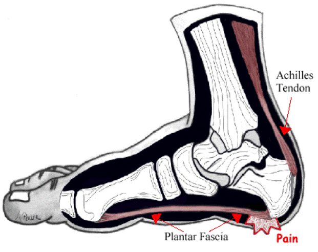
Arch pain typically is the term used to describe pain under the arch of the foot. Arch pain indicates inflammation of the tissues within the midfoot and is most commonly caused by plantar fasciitis. Plantar fasciitis is inflammation of the fibrous band of tissue that connects the heel to the toes. Arch pain is most commonly found early in the morning due to the plantar fascia becoming contracted and tight during sleep. Walking or standing for long periods of time can also aggravate the plantar fascia, causing it to become inflamed and irritated. Treatment options include orthotics, anti-inflammatory medications and stretching exercises.
Causes
There are many causes for a high arch (cavus) foot. In the United States, the most common cause for a high arch foot is a form of muscular dystrophy called hereditary sensorimotor neuropathy. Most people recognize this by the more commonly used name of Charcot Marie Tooth disease (CMT). This is a disease of the muscles and the nerves of the legs, and occasionally of the hands, in which certain muscles weaken while others retain their strength. The condition is transmitted as an autosomal dominant condition. This means that 50% of the offspring will statistically inherit the disorder. This is, however, just a statistic. In some families, all the children develop the condition while in others, none inherit it.
Symptoms
The foot of a newborn with congenital vertical talus typically has a convex rocker-bottom shape. This is sometimes combined with an actual fold in the middle of the foot. The rare person who is diagnosed at an older age often has a "peg-leg" gait, poor balance and heavy calluses on the soles where the arch would normally be. If a child with congenital vertical talus has a genetic disorder, additional symptoms often are seen in other parts of the body.
Diagnosis
The doctor will examine your feet for foot flexibility and range of motion and feel for any tenderness or bony abnormalities. Depending on the results of this physical examination, foot X-rays may be recommended. X-rays are always performed in a young child with rigid flatfeet and in an adult with acquired flatfeet due to trauma.
Non Surgical Treatment
Just as there are many different causes of flat feet, there are also many different treatment options. The most important aspect of treatment is determining the exact type or underlying cause of flat feet that you have. Foot and ankle specialists can determine this through thorough clinical examination and special imaging studies (e.g., x-rays, computed tomography, and/or magnetic resonance imaging). Conservative treatment is effective in the vast majority of flat foot cases, and consists of things such as insoles, splints, manipulation, or casting. Surgery is required much less frequently, and is reserved only for some of the severe types of flat foot that do not respond to conservative therapy.

Surgical Treatment
Patients with adult acquired flatfoot are advised to discuss thoroughly the benefits vs. risks of all surgical options. Most procedures have long-term recovery mandating that the correct procedure be utilized to give the best long-term benefit. Most flatfoot surgical procedures require six to twelve weeks of cast immobilization. Joint fusion procedures require eight weeks of non-weightbearing on the operated foot, meaning you will be on crutches for two months. The bottom line is: Make sure all of your non-surgical options have been covered before considering surgery. Your primary goals with any treatment are to eliminate pain and improve mobility. In many cases, with the properly designed foot orthosis or ankle brace, these goals can be achieved without surgical intervention.
Stretching Exercises
Stretching your calf and Achilles tendon may also help as a tight Achilles can make your foot overpronate. To stretch your calf and Achilles tendon, step forwards with your left leg and bend it, with your right leg straight and both feet pointing forwards. Push your right heel into the ground while keeping your right leg straight; you should feel the stretch at the back of your right leg, below the knee. Hold the stretch for 15 to 30 seconds and repeat with the opposite leg. Repeat the stretch two to four times on each leg, and repeat the overall exercise three to four times a day.
Arch pain typically is the term used to describe pain under the arch of the foot. Arch pain indicates inflammation of the tissues within the midfoot and is most commonly caused by plantar fasciitis. Plantar fasciitis is inflammation of the fibrous band of tissue that connects the heel to the toes. Arch pain is most commonly found early in the morning due to the plantar fascia becoming contracted and tight during sleep. Walking or standing for long periods of time can also aggravate the plantar fascia, causing it to become inflamed and irritated. Treatment options include orthotics, anti-inflammatory medications and stretching exercises.
Causes
There are many causes for a high arch (cavus) foot. In the United States, the most common cause for a high arch foot is a form of muscular dystrophy called hereditary sensorimotor neuropathy. Most people recognize this by the more commonly used name of Charcot Marie Tooth disease (CMT). This is a disease of the muscles and the nerves of the legs, and occasionally of the hands, in which certain muscles weaken while others retain their strength. The condition is transmitted as an autosomal dominant condition. This means that 50% of the offspring will statistically inherit the disorder. This is, however, just a statistic. In some families, all the children develop the condition while in others, none inherit it.
Symptoms
The foot of a newborn with congenital vertical talus typically has a convex rocker-bottom shape. This is sometimes combined with an actual fold in the middle of the foot. The rare person who is diagnosed at an older age often has a "peg-leg" gait, poor balance and heavy calluses on the soles where the arch would normally be. If a child with congenital vertical talus has a genetic disorder, additional symptoms often are seen in other parts of the body.
Diagnosis
The doctor will examine your feet for foot flexibility and range of motion and feel for any tenderness or bony abnormalities. Depending on the results of this physical examination, foot X-rays may be recommended. X-rays are always performed in a young child with rigid flatfeet and in an adult with acquired flatfeet due to trauma.
Non Surgical Treatment
Just as there are many different causes of flat feet, there are also many different treatment options. The most important aspect of treatment is determining the exact type or underlying cause of flat feet that you have. Foot and ankle specialists can determine this through thorough clinical examination and special imaging studies (e.g., x-rays, computed tomography, and/or magnetic resonance imaging). Conservative treatment is effective in the vast majority of flat foot cases, and consists of things such as insoles, splints, manipulation, or casting. Surgery is required much less frequently, and is reserved only for some of the severe types of flat foot that do not respond to conservative therapy.
Surgical Treatment
Patients with adult acquired flatfoot are advised to discuss thoroughly the benefits vs. risks of all surgical options. Most procedures have long-term recovery mandating that the correct procedure be utilized to give the best long-term benefit. Most flatfoot surgical procedures require six to twelve weeks of cast immobilization. Joint fusion procedures require eight weeks of non-weightbearing on the operated foot, meaning you will be on crutches for two months. The bottom line is: Make sure all of your non-surgical options have been covered before considering surgery. Your primary goals with any treatment are to eliminate pain and improve mobility. In many cases, with the properly designed foot orthosis or ankle brace, these goals can be achieved without surgical intervention.
Stretching Exercises
Stretching your calf and Achilles tendon may also help as a tight Achilles can make your foot overpronate. To stretch your calf and Achilles tendon, step forwards with your left leg and bend it, with your right leg straight and both feet pointing forwards. Push your right heel into the ground while keeping your right leg straight; you should feel the stretch at the back of your right leg, below the knee. Hold the stretch for 15 to 30 seconds and repeat with the opposite leg. Repeat the stretch two to four times on each leg, and repeat the overall exercise three to four times a day.
 RSS Feed
